Review


rabbit polyclonal anti flotilin  (Novus Biologicals)


Bioz Verified Symbol Novus Biologicals is a verified supplier

Bioz Manufacturer Symbol Novus Biologicals manufactures this product  

  • Logo
  • About
  • News
  • Press Release
  • Team
  • Advisors
  • Partners
  • Contact
  • Bioz Stars
  • Bioz vStars
  • 93

    Structured Review

    Novus Biologicals rabbit polyclonal anti flotilin
    Rabbit Polyclonal Anti Flotilin, supplied by Novus Biologicals, used in various techniques. Bioz Stars score: 93/100, based on 3 PubMed citations. ZERO BIAS - scores, article reviews, protocol conditions and more
    https://www.bioz.com/product/flotilin+1/pm41264708-591-238-243?v=Novus+Biologicals
    Average 93 stars, based on 3 article reviews
    rabbit polyclonal anti flotilin - by Bioz Stars, 2026-07
    93/100 stars

    Images


    Similar Products

    93
    Novus Biologicals rabbit polyclonal anti flotilin
    Rabbit Polyclonal Anti Flotilin, supplied by Novus Biologicals, used in various techniques. Bioz Stars score: 93/100, based on 1 PubMed citations. ZERO BIAS - scores, article reviews, protocol conditions and more
    https://www.bioz.com/product/flotilin+1/pm41264708-591-238-243?v=Novus+Biologicals
    Average 93 stars, based on 1 article reviews
    rabbit polyclonal anti flotilin - by Bioz Stars, 2026-07
    93/100 stars
      Buy from Supplier

    93
    Novus Biologicals flotilin 1
    Flotilin 1, supplied by Novus Biologicals, used in various techniques. Bioz Stars score: 93/100, based on 1 PubMed citations. ZERO BIAS - scores, article reviews, protocol conditions and more
    https://www.bioz.com/product/flotilin+1/10__1016_slash_j__snb__2024__136939-136-29-33?v=Novus+Biologicals
    Average 93 stars, based on 1 article reviews
    flotilin 1 - by Bioz Stars, 2026-07
    93/100 stars
      Buy from Supplier

    95
    Cell Signaling Technology Inc anti flotilin
    Anti Flotilin, supplied by Cell Signaling Technology Inc, used in various techniques. Bioz Stars score: 95/100, based on 1 PubMed citations. ZERO BIAS - scores, article reviews, protocol conditions and more
    https://www.bioz.com/product/flotilin+1/pm39875894-64-4-8?v=Cell+Signaling+Technology+Inc
    Average 95 stars, based on 1 article reviews
    anti flotilin - by Bioz Stars, 2026-07
    95/100 stars
      Buy from Supplier

    86
    Cell Signaling Technology Inc anti flotilin 1
    Anti Flotilin 1, supplied by Cell Signaling Technology Inc, used in various techniques. Bioz Stars score: 86/100, based on 1 PubMed citations. ZERO BIAS - scores, article reviews, protocol conditions and more
    https://www.bioz.com/product/flotilin+1/pm39262778-147-30-32?v=Cell+Signaling+Technology+Inc
    Average 86 stars, based on 1 article reviews
    anti flotilin 1 - by Bioz Stars, 2026-07
    86/100 stars
      Buy from Supplier

    95
    Cell Signaling Technology Inc flotilin 1
    Flotilin 1, supplied by Cell Signaling Technology Inc, used in various techniques. Bioz Stars score: 95/100, based on 1 PubMed citations. ZERO BIAS - scores, article reviews, protocol conditions and more
    https://www.bioz.com/product/flotilin+1/pm39210346-257-37-40?v=Cell+Signaling+Technology+Inc
    Average 95 stars, based on 1 article reviews
    flotilin 1 - by Bioz Stars, 2026-07
    95/100 stars
      Buy from Supplier

    95
    Santa Cruz Biotechnology anti flotilin
    Anti Flotilin, supplied by Santa Cruz Biotechnology, used in various techniques. Bioz Stars score: 95/100, based on 1 PubMed citations. ZERO BIAS - scores, article reviews, protocol conditions and more
    https://www.bioz.com/product/flotilin+1/pm38999977-341-73-79?v=Santa+Cruz+Biotechnology
    Average 95 stars, based on 1 article reviews
    anti flotilin - by Bioz Stars, 2026-07
    95/100 stars
      Buy from Supplier

    90
    Becton Dickinson anti–flotilin-1
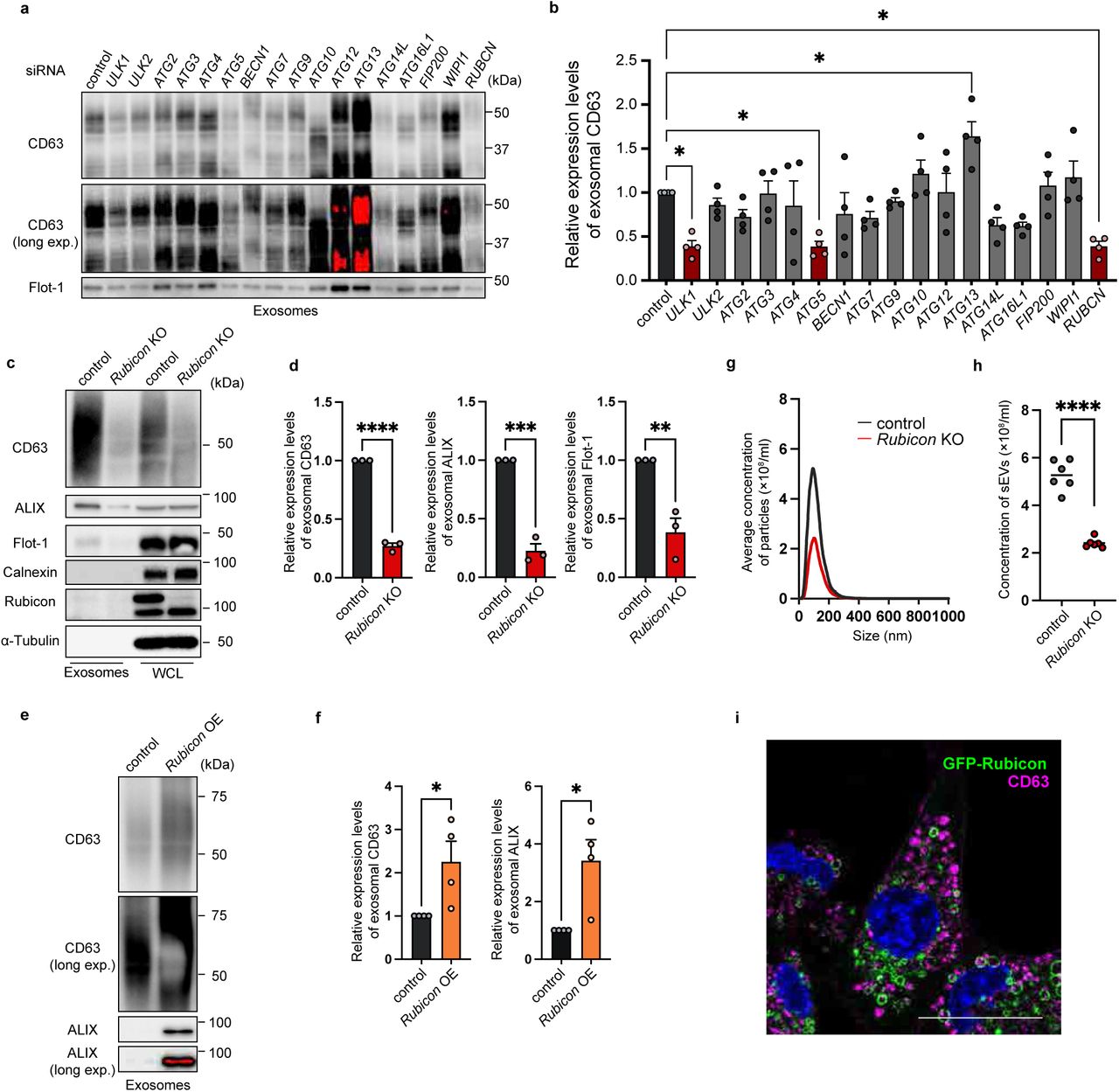
    (a) Immunoblotting of CD63 and <t>Flotilin-1</t> (Flot-1) in the exosome fractions isolated from the culture medium of human mesenchymal stem cells (hMSCs) with knockdown of the indicated genes. The loading amount for each sample was set as follows: half of the exosome sample collected from 1 ml of culture medium. (b) Quantification of the exosomal CD63 levels in the hMSCs with knockdown of the genes indicated in (a). Bars represent means ± SEM. n = 4 independent experiments. *p < 0.05 (each vs control) by one-way ANOVA followed by Dunnett’s test. (c) Immunoblotting of the indicated proteins in the exosome fractions and the WCLs obtained from control and Rubicon -knockout (KO) mouse embryo fibroblasts (MEFs). The loading amount for each sample was set as follows: half of the exosome sample collected from 1 ml of culture medium, and 6 µg for the WCL. (d) Quantification of the exosomal levels of CD63, ALIX, and Flot-1 in (c). Bars represent means ± SEM. n = 3 independent experiments. **p < 0.01, ***p < 0.001, ****p < 0.0001 by a two-tailed Student’s t-test. (e) Immunoblotting of CD63 and ALIX in the exosome fractions isolated from the cultured medium of control and Rubicon-overexpressing (OE) MEFs. The loading amount for each sample was set as follows: half of the exosome sample collected from 1 ml of culture medium. (f) Quantification of the exosomal CD63 and ALIX levels in (e). Bars represent means ± SEM. n = 4 independent experiments. *p < 0.05 by a two-tailed Student’s t-test. (g) Nanoparticle tracking analysis (NTA) of the extracellular vesicles (EVs) purified from the cultured medium of control or Rubicon -KO MEFs using a phosphatidylserine affinity magnetic resin (PS-affinity kit). (h) Quantification of the total particle concentration of the EVs in (g). Bars represent means. n = 6 independent experiments. ****p < 0.0001 by a two-tailed Student’s t-test. (i) Immunofluorescent images of CD63 (magenta) and DAPI (blue) in MEFs stably expressing GFP-Rubicon treated with 0.5 μM Apilimod for 1 hour. Scale bars, 50 μm.
    Anti–Flotilin 1, supplied by Becton Dickinson, used in various techniques. Bioz Stars score: 90/100, based on 1 PubMed citations. ZERO BIAS - scores, article reviews, protocol conditions and more
    https://www.bioz.com/product/flotilin+1/bio_rxiv__2024__05__08__593233-185-19-20?v=Becton+Dickinson
    Average 90 stars, based on 1 article reviews
    anti–flotilin-1 - by Bioz Stars, 2026-07
    90/100 stars
      Buy from Supplier

    Image Search Results


    (a) Immunoblotting of CD63 and Flotilin-1 (Flot-1) in the exosome fractions isolated from the culture medium of human mesenchymal stem cells (hMSCs) with knockdown of the indicated genes. The loading amount for each sample was set as follows: half of the exosome sample collected from 1 ml of culture medium. (b) Quantification of the exosomal CD63 levels in the hMSCs with knockdown of the genes indicated in (a). Bars represent means ± SEM. n = 4 independent experiments. *p < 0.05 (each vs control) by one-way ANOVA followed by Dunnett’s test. (c) Immunoblotting of the indicated proteins in the exosome fractions and the WCLs obtained from control and Rubicon -knockout (KO) mouse embryo fibroblasts (MEFs). The loading amount for each sample was set as follows: half of the exosome sample collected from 1 ml of culture medium, and 6 µg for the WCL. (d) Quantification of the exosomal levels of CD63, ALIX, and Flot-1 in (c). Bars represent means ± SEM. n = 3 independent experiments. **p < 0.01, ***p < 0.001, ****p < 0.0001 by a two-tailed Student’s t-test. (e) Immunoblotting of CD63 and ALIX in the exosome fractions isolated from the cultured medium of control and Rubicon-overexpressing (OE) MEFs. The loading amount for each sample was set as follows: half of the exosome sample collected from 1 ml of culture medium. (f) Quantification of the exosomal CD63 and ALIX levels in (e). Bars represent means ± SEM. n = 4 independent experiments. *p < 0.05 by a two-tailed Student’s t-test. (g) Nanoparticle tracking analysis (NTA) of the extracellular vesicles (EVs) purified from the cultured medium of control or Rubicon -KO MEFs using a phosphatidylserine affinity magnetic resin (PS-affinity kit). (h) Quantification of the total particle concentration of the EVs in (g). Bars represent means. n = 6 independent experiments. ****p < 0.0001 by a two-tailed Student’s t-test. (i) Immunofluorescent images of CD63 (magenta) and DAPI (blue) in MEFs stably expressing GFP-Rubicon treated with 0.5 μM Apilimod for 1 hour. Scale bars, 50 μm.

    Journal: bioRxiv

    Article Title: The Rubicon-WIPI axis regulates exosome biogenesis during aging

    doi: 10.1101/2024.05.08.593233

    Figure Lengend Snippet: (a) Immunoblotting of CD63 and Flotilin-1 (Flot-1) in the exosome fractions isolated from the culture medium of human mesenchymal stem cells (hMSCs) with knockdown of the indicated genes. The loading amount for each sample was set as follows: half of the exosome sample collected from 1 ml of culture medium. (b) Quantification of the exosomal CD63 levels in the hMSCs with knockdown of the genes indicated in (a). Bars represent means ± SEM. n = 4 independent experiments. *p < 0.05 (each vs control) by one-way ANOVA followed by Dunnett’s test. (c) Immunoblotting of the indicated proteins in the exosome fractions and the WCLs obtained from control and Rubicon -knockout (KO) mouse embryo fibroblasts (MEFs). The loading amount for each sample was set as follows: half of the exosome sample collected from 1 ml of culture medium, and 6 µg for the WCL. (d) Quantification of the exosomal levels of CD63, ALIX, and Flot-1 in (c). Bars represent means ± SEM. n = 3 independent experiments. **p < 0.01, ***p < 0.001, ****p < 0.0001 by a two-tailed Student’s t-test. (e) Immunoblotting of CD63 and ALIX in the exosome fractions isolated from the cultured medium of control and Rubicon-overexpressing (OE) MEFs. The loading amount for each sample was set as follows: half of the exosome sample collected from 1 ml of culture medium. (f) Quantification of the exosomal CD63 and ALIX levels in (e). Bars represent means ± SEM. n = 4 independent experiments. *p < 0.05 by a two-tailed Student’s t-test. (g) Nanoparticle tracking analysis (NTA) of the extracellular vesicles (EVs) purified from the cultured medium of control or Rubicon -KO MEFs using a phosphatidylserine affinity magnetic resin (PS-affinity kit). (h) Quantification of the total particle concentration of the EVs in (g). Bars represent means. n = 6 independent experiments. ****p < 0.0001 by a two-tailed Student’s t-test. (i) Immunofluorescent images of CD63 (magenta) and DAPI (blue) in MEFs stably expressing GFP-Rubicon treated with 0.5 μM Apilimod for 1 hour. Scale bars, 50 μm.

    Article Snippet: The following antibodies were used for western blotting at the indicated dilutions: anti-CD63 (MBL, D263-3, 1:5000; Abcam, ab134045, 1:2000), anti–Flotilin-1 (BD Biosciences, 610821, 1:10000), anti-ALIX (BioLegend, 634502, 1:1000), anti-Calnexin (Sigma-Aldrich, HPA009433, 1:1000), anti-Rubicon (Cell Signaling Technology, 8465, 1:2000), anti–α-Tubulin (PM054, MBL, 1:20000), anti-WIPI2 (Sigma-Aldrich, SAB4200400, 1:2000), anti-HRS (GeneTex, GTX101718, 1:1000), anti-VPS4 (Sigma-Aldrich, SAB4200025, 1:1000), anti-GFP (Cell Signaling Technology, 2555, 1:1000), anti-p21 (Abcam, ab109199, 1:1000), anti-γH2AX (Abcam, ab2893, 1:1000), anti-p16 INK4a (IBL, 11104, l g/ml), anti-LC3 (MBL, PM036, 1:2000), anti-Atg12 (Cell Signaling Technology, 2011S, 1:1000), anti-FIP200 (Proteintech, 17250-1-AP, 1:1000), anti-Atg2A (MBL, PD041, 1:1000), anti-Atg2B (Sigma-Aldrich, HPA019665, 1:1000), anti-Atg14 (Cell Signaling Technology, 96752S, 1:1000), anti–Beclin-1 (Cell Signaling Technology, 3738S, 1:1000), anti-UVRAG (MBL, M160-3, 1:2000), anti-ATG16L1 (Cell Signaling Technology, 8089, 1:1000), anti-PIK3C3 (Cell Signaling Technology, 3811, 1:1000), anti-WIPI1 (Sigma-Aldrich, W4769, 1:1000), anti-WIPI3 (Thermo Fisher Scientific, PA5-50864, 1:1000), anti-WIPI4 (Proteintech, 19194-1-AP, 1:1000), anti–β-Actin (MBL, M177-3.

    Techniques: Western Blot, Isolation, Knock-Out, Two Tailed Test, Cell Culture, Purification, Concentration Assay, Stable Transfection, Expressing